Common Diseases & Conditions in Pets F-G
False pregnancy (also known as pseudopregnancy) is very common in canines but not so in cats. It is caused by a hormone inbalance. The suspect hormone is progesterone, a hormone needed for maintenance of pregnancy but in excess can also lead to pyometra.
False pregnancies almost always occur about a month after the intact female was in heat; yet she had not mated with a male dog. Hormonal changes in the dogs body cause about 100% of all the clinical signs seen.
Most people presenting a dog for false pregnancy are flabbergasted to say the least. They swear up and down that their dog did not “get caught”. I usually told them that this happens and it isn’t the clients fault! The clinical signs associated are those very similar to a female about ready to deliver her pups! The dog may: have breast enlargement and often start to lactate (very common), start to pace and become anxious, make a nesting place for her “phantom pups” and actually go into labor in some rare instances!
Lab work includes a CBC and Chemistry profile plus a vaginal smear to be stained and examined under the microscope. Radiographs may be taken to MAKE SURE MISTAKES are not made. People reading this would never believe the things I have heard or seen in my 33 year long career! I have had people swear to me on a stack of Bibles that the dog was not impregnated. Close to 2 months from the time the dog was in heat, the animal is presented for (at least it looked like it from the history) false pregnancy. Low and behold, a lateral radiograph showed a bunch of fetal skeletons in the uterus. You get the point! Always make sure the dog is not pregnant!
Diagnosis is easily made by the history obtained and a dog that was in estrous 1-2 months prior to presentation. Lab Work and radiographs will rule out pregnancies and the like.
There is no specific therapy for pseudopregnancy as the clinical signs will subside over time. If it is a breeding animal, the dog is fully capable of being mated in the future.
If the dog is not to be bred, it is ultra important that all dogs going through a false pregnancy be spayed. Going through false pregnancies increases the risk of developing breast cancer in the dog. Progesterone excess (one of the causes) can cause a female to develop pyometra; a severe uterine infection requiring an emergency ovariohysterectomy.
The prognosis for false pregnancy is excellent. Prevention of future episodes of this condition and other gynecological problems can be totally avoided by having the animal spayed.
Feline Immunodeficiency Virus (FIV) is a debilitating virus of cats caused by a retrovirus. It is in the same class as Feline Leukemia.
The FIV attacks the immune system of the cat weakening it in the process. This makes the animal much more susceptible to any future infection. It is very commonly seen in male tom cats plus cats that live outdoors. The mode of transmission is primarily through bite wounds from cat to cat. Retroviruses are species specific. The cat virus does not cause disease in dogs or humans.
The cat goes through several stages of disease: acute, subacute and chronic. Throughout all of these stages, the immune system gets weakened even further. By the time the cat is in the chronic phase, bacteria and or viruses that rarely cause trouble in a healthy cat, can be fatal. The immune system has literally been torn to shreds.
The clinical signs associated with FIV are varied. The entire immune system is deficient so all body systems can be affected by the virus. One of the most common signs seen in the cat are: a poor coat, anorexia and weight loss. These are general signs. A specific sign seen in at least half of all FIV cats is the presence of oral lesions. Severely infected gums (gingivitis), mouth (stomatitis) and severe dental disease are seen. Animals often lose weight due to the persistent diarrhea that exists. Chronic wasting and respiratory infections are seen as well.
A CBC and Chemistry profile will show altered values due to the organs affected by FIV. A conclusive test is available and most veterinarians use a combo Idexx® FIV/FeLV Snap Test. The clinical signs of both diseases are similar so it is wise to run both tests at the same time. There are some issues though:
1. The snap test detects antibodies to FIV. Young kittens may also test positive since their mother passes short term passive immunity to them via nursing. Retest every 60 days till they are about 6 months of age. Cats that are vaccinated against FIV will test positive since vaccine antibodies (at the moment) can not be differentiated from actual infection antibodies.
2. A negative snap test means there are no antibodies present and can be assumed to be free of the disease…except in those cats just exposed to the virus and tested, there will be no antibodies. Most diseases require at least 2 months to develop an effective humoral immune response.
No lab test is ever 100% effective one way or another.
Diagnosis is made by the associated batch of clinical signs that can be present plus by the history of the cat being male and living outdoors or completely feral. Even though their are interpretive problems with the snap test, it is highly effective in leading to a positive diagnosis when combined with other physical findings.
There is no actual direct treatment for FIV infections. If secondary infections are present, those need to be treated and fluid replacement and calories provided via Nutrical® or any other high calorie food. The goal in positive cats is to allow the animal to have a quality of life without exposing other cats to the disease. Sick cats should always be kept indoors and away from other animals. What if there are other cats in the household? It is risky. They can all be tested and vaccinated but no vaccine is perfect and there is a chance of exposure. That is a risk an owner will have to decide whether they are willing to try that approach. Sick cats have a completely demoralized immune system and can not handle any stress. Try to keep the environment as stress free as possible.
The prognosis of the disease varies with the organ systems that are effected plus the severity of the immune system degregation. If an FIV cat stabilizes it can live for many years as long as periodic medical exams and lab work are performed to assess bodily functions. Any other infectious disease or condition must be immediately treated. It is preferable not to introduce any other cats to the household at this time. The risk of exposure is too high. Prognosis becomes poor when an animal starts to lose tremendous amounts of weight over a short time and a worsening of its condition.
Prevention is the key with many diseases in the cat. Testing, testing and more testing. That is it in a nutshell. Without screening, it is impossible to know which cat is positive and which is negative. Screen all cats new to the household via adoption or a cat found as a stray. Many veterinarians test ANY sick cat that enters the practice. As I preached to my Ohio clients, keep the cat indoors!! When it comes to FIV, that is very good advice.

Feline Herpes Virus (FHV-1) is also known as Feline Viral Rhinotracheitis. It causes severe respiratory infections in cats; particularly in kittens.
Feline Herpes Virus is a tricky little bug. Like all Herpes viruses, a patient is never free of the virus. They may recover from the illness but the virus is always present. Herpes virus is a latent virus. It hides in neutrophils and other blood cells and effectively evades the immune system. Once the cat is stressed from some exogenous cause, the virus leaves its hiding place and pounces on the cat causing a repeat of respiratory signs.
Feline Herpes Virus is transmitted by direct contact from cat to cat by any nasal or eye secretion. Bedding and food bowls can be a source of contamination to susceptible cats. It is seen most commonly in places where there are large numbers of cats in close quarters such as shelters and catteries. Young kittens are at a higher risk since they have an immature immune system. The tricky part in cat exposure is the carrier cat. Those cats have a latent infection. They are not clinically ill but can easily transmit the virus to a susceptible cat.
Clinical signs in cats are always respiratory. Intense sneezing, coughing, nasal discharges, runny eyes and infected conjunctiva are all part of the disease process. A severe head cold! Animals will be anorexic and, like most sick cats, will just sit in one place for hours with their heads held down.
Veterinarians will initially draw a CBC and Chemistry profile to check bodily functions. Being a virus, herpes cats will often have a drop in their white cell count (leukopenia). Swabs of the nasal secretions or conjunctiva will be sent to the lab for culture and hence a definitive diagnosis. Chest films may be taken in case of severe respiratory infections.
Diagnosis of Feline Herpes Virus is made through a good history and physical exam. Many other viruses can mimic the FHV-1 virus. Conclusive diagnosis is made by culturing the virus in the lab.
There is no specific treatment for Feline Herpes Virus. Antibiotics are usually prescribed to prevent secondary bacterial infections. Nebulization therapy is also effective in clearing the respiratory tract of secretions making it easier for the cat to breathe. Topical ophthalmic antibiotics are usually prescribed to treat the bilateral conjunctivitis. L-Lysine (Viralys®) has been found to minimize the clinical signs associated with herpes virus by slowing down the intracellular replication of the herpes virus. L-Lysine is a nutritional supplement and comes in gel form for easy administration to the cat.
The prognosis for most Feline Herpes Virus patients is very good. Providing a stress free environment for the cat and high quality nutrition goes a long way in helping improve the life of the cat. It is a latent virus and can cause issues in the future.
Prevention is the key. Feline Viral Rhinotracheitis is one of the three diseases commonly vaccinated for in cats beginning at six weeks of age. No vaccine is perfect but this one is a good bet for all cats. The kitten needs to go through a complete series of shots than boostered once annually to maintain effective antibody levels to the disease.
Feline Infectious Peritonitis (FIP) is a severe, debilitating disease in cats. Young cats with an immature immune system are most commonly infected with the virus. FIP is a member of the cornona virus family. Corona viruses usually are associated with digestive clinical signs in the dog and cat but a cornona virus serotype was found to cause the clinical signs of FIP in the cat.
FIP is extremely contagious and passed easily from one cat to another from bodily fluids and or secretions. The disease exists in two forms:
1. Wet form: Usually seen in young kittens, this form is characterized by the accumulation of fluid within the abdominal cavity or thorax or both. Clinical signs will be related to the presence of fluids in these cavities.
2. Dry form: In this form, granulomas (scar tissue) are formed in various organs particularly the liver, lungs and spleen. Clinical signs are based on the location of the lesions. This form can be challanging to diagnose.
Clinical signs associated with the wet form of FIP is the presence of a distended abdomen and or labored breathing due to the presence of free fluid in the pleural space or abdominal cavity. Cats will often have respiratory signs or diarrhea that are non responsive to antibiotics. Cats become anorexic and lose weight rapidly. The dry form of the disease can be insidious. The cat may harbor the virus without any signs for years. They will act like nothing is wrong. When stressed the granulomas take their toll and cats will develop signs of liver disease such as weight loss and jaundice. The major problem with cats is that they can be deathly ill, and from an outward appearance, they feel like they have the world in their paws (they usually do anyway!). Stress comes along, than they crash. This makes it much harder to treat them because they are so far along when diagnosed.
An FIP ELISA (Enzyme Linked Immunosorbent Assay Test) exists but unfortunately it is unable to differentiate the intestinal version of corona virus from the FIP version of corona virus. All a positive test states is that the cat had been exposed to one or the other but will not tell you which one. A negative test can assume the cat is negative for both. The problem with that is, if the cat has just developed clinical signs it may be too early for an antibody response to be picked up hence giving a false negative. A CBC and Chemistry profile are always done plus a urinalysis to monitor bodily functions for an source of disease.
Diagnosis of the wet form is made by a history of exposure plus associated clinical signs. Tapping the abdomen will almost always produce a straw colored liquid that is sticky between the fingers. This is due to the protein content of the liquid. The dry form is extremely difficult to diagnose because of the varied signs seen that may mimic other disease processes. Even when suspected, there is no effective test to conclusively diagnose it. Therefore, FIP is usually a tentative diagnosis. It could be confirmed on tissue sample submission but by that time the cat is too weak and a poor anesthetic risk to do the procedure. Many cats are diagnosed post mortem.
There is no direct treatment that will neutralize the FIP virus. In the wet form, clinical signs develop so rapidly that most cats succumb within a week or two or less. Once the dry form manifests itself, it too is difficult to treat with supportive care. Supportive care can be attempted. This includes: fluid and calorie support and appetite stimulation with mirtazapine. Vitamins may also be given. In the end, you are just buying time and most cats are euthanized.
The prognosis for FIP cases is usually poor. There is no antiviral therapy and supportive care is iffy at best. There is a FIP vaccine that came out in the mid 1990’s but its level of effectivity was initially poor. Only about 50%-60% of cats developed any benefit from the vaccine. I used it in my Ohio practice but was not impressed. It was an intranasal vaccine. I do not recommend its use.
Feline Leukemia is a severe viral disease in cats that plays havoc with its immune system. It is caused by a retrovirus and is often known as the AIDS virus of cats. Back in the early 1980’s and before the FeLV vaccine, I would diagnose at least 5 cats every day with it. It was THAT common before vaccination became popular and highly effective.
Feline Leukemia (FeLV) is extremely contagious and is transmitted by bite wounds, nasal and fecal secretions and by sharing infected food & water dishes and litter boxes. It is not common in indoor cats but most commonly seen in catteries, shelters or feral cats. The sad thing is, is that the virus can be transmitted in utero to a queens unborn kittens. I have watched litters of kittens die after trying to get a start on life but only to succumb to the disease because the mother was a carrier. It is this carrier state that is dangerous. The cat will look and act clinically normal but be able to transmit the disease to other cats. FeLV is not transmissable to people and dogs. Once diagnosed with the disease, most cats almost never live more than 2-3 years longer. The disease is most commonly seen in young kittens up to about the age of 6. Older cats when exposed may ward off the virus due to a healthy immune response.
Clinical signs associated with FeLV are all related to the immune system decline seen in all cats. In some forms, the cat literally melts in front of your eyes. Signs of extreme weight loss and dehyration are common. Anemia and jaundice are common. The anemia is noticeable by the pale gums of the mouth and jaundice always starts around the soft palate of the oral cavity. Some animals may have persistent respiratory or digestive signs. Animals may have a persistent fever and enlarged lymph nodes. Some will have tumors related to the condition. Lots of cats show ZERO clinical signs and they are dangerous carriers of the disease. Once stressed with respiratory infections or facsimile, they too can demonstrate clinical signs. A pregnant queen can can be suspect of carrying the disease if her kittens soon die. FeLV is at the top of the list in so called “fading kitten syndrome diseases”.
All sick cats should undergo a CBC and Chemistry profile for a minimal clinical database. Usually, a combo Idexx® FeLV/FIV Snap Test is performed. FIV mimics FeLV in many of its clinical signs, so it makes sense to run the combo test.
Diagnosis is usually made in a young cat demonstrating clinical signs of FeLV. These cats have not been vaccinated against the disease. Association with other cats in catteries, shelters or multi-cat households, where the disease is more common, is an important historical finding. The FeLV ELISA test confirms the diagnosis.
There is no direct treatment to eliminate the Feline Leukemia Virus. In homes with one infected cat, supportive care can be commenced which entails a high caloric diet and a multi-vitamin gel such as Nutrical® should always be offered. A handy trick is to mix Nutrical® together with Hill’s® Prescription a/d food. This combination really packs a punch and definitely helps the cat.
The tricky question becomes what to do about a positive FeLV cat that is living with other cats in the household? On one hand, it is dangerous to have a positive cat around other healthy cats since the virus can be transmitted to the other cats. On the other hand, the positive cat may have been healthy and around the other cats for a long period of time. It is very possible and highly likely that the other cats were exposed to the virus and were able to fight it off due to a healthy immune response. What to do varies with each owner. Some owners will wish the positive cat put down while others want to provide supportive care. Ideally, the positive cat should be kept separate from the other cats. All of the remaining cats should be tested for the virus and if negative should all be vaccinated against FeLV. If a cat tests positive and is healthy, this is not a death sentence. That cat should be tested every 1-2 months and if the veterinarian gets two consecutive negative tests, the animal should be vaccinated for FeLV. Cats are fully capable of fighting off an infection.
Even with supportive care, the long term prognosis for FeLV cats is poor. Most cats rarely live longer than 3 years after the condition is diagnosed. It is heartbreaking to see these animals decline. Over the years, it has made practicing veterinary medicine very difficult at times.
The best approach is prevention. All cats that are taken home should be vaccinated and tested. Keep all cats indoors and away from any sick cat. If your one cat is vaccinated against FeLV and you want to get another cat, make sure a FeLV test has been performed and get the cat vaccinated. If you find a stray cat or buy a cat and you are unsure if the animal has been tested, DO NOT TAKE THE CAT HOME IMMEDIATELY. Take it to your veterinarian and get it tested. If negative than you can introduce the new cat. No vaccine is purrfect but it is known that a vaccinated cat is a safer, healthier cat in case it is every challenged by a positive FeLV animal.

Everybody knows that fish hooks are used for fishing. Bait is applied to the hook and people catch fish! This is great but not so when an animal gets caught in one or all of its nasty barbs.
Most cases of animals getting entangled in fish hooks are at camping sites, lakes, oceans or anywhere people go fishing. Many of the fish hooks have a fishy/meaty smell that attracts both cats and dogs. Fish hooks also have other shiny components that also make them irresistible to cats. Felines love any and I mean any shiny object. Fish hooks can be embedded anywhere on the body. Fish hooks may also be swallowed. This can be quite dangerous.
Fish hooks are many times found embedded: in the paws of the animal, the tongue, the cheeks of the mouth and the nostrils most frequently.
Many times owners do not notice anything wrong until they see fish line hanging off of a dog or cats body. Because of this, veterinarians will run a CBC to mainly check the white cell count for signs of infection (a left shift neutrophilia).
Diagnosis is made by finding a fish hook embedded anywhere on the animal’s body.
Treatment is effective removal of the fish hook. This is always done under light sedation. Usually the barb is pushed threw the tissue, the sharp barb is cut off and the remainder of the hook is pulled out the way it went in. Forcing it out without cutting the barb will do more harm than good. Animals may also swallow the fish hook. If this is in the oral cavity, it is easily removed. If it is in the esophagus or stomach it may need to be removed with an endoscope or surgery. If the hook is in the intestinal tract surgery may be necessary. Antibiotics are always prescribed for infections and pain killers may be prescribed for a short period.
Prognosis for fish hook altercations is excellent. Prevention of the problem is best. Keep all tackle boxes and bait supplies closed and away from prying dogs and cats. If warranted, keep animals away from the fishing site. This is complete avoidance of anything bad happening.


Fleas cause total discomfort in dogs and cats. Most of that discomfort revolves around the severe allergy developed to the flea bite. Rarely do people ever think that an animal can become anemic over time to fleas. A flea is a little blood sucker and drains your pet of blood one drop at a time. If the animal is extremely small and young plus severely infested with fleas, anemia can happen.
A small dog or cat has a very low blood volume. If it is covered from head to toe with fleas, blood depletion will soon happen. Making matters worse, fleas transmit tapeworms to dogs and cats. Those parasites can further drain an animal of any blood or nutrition. Flea anemia can easily cause the death of a small, young puppy or kitten.
Pale mucous membranes and pale pads of the paws are noticed. More systemic clinical signs may be seen due to the anemia. The respiratory and heart rate will be elevated and the animal may tire often. Seeing this in active puppies and kittens is abnormal. Young animals are very active. Pets parasitized with tapeworms may also pass those tape segments or other intestinal parasites.
A CBC is the lab test of choice to check for anemia. Levels of hemoglobin and red cells will be lower. The hematocrit will also be lower. Smears will be made to determine the type of anemia. Fecal samples will be checked for intestinal parasites.
Diagnosis is made by the presence of large flea populations on the dog or cat plus the clinical signs of anemia.
In severe cases of anemia, a pet may require a blood transfusion. In most cases hematinics are administered to the animal. Nutrical® contains extra iron and other nutrients to help an animal. Most dogs have a regenerative anemia so will respond to this treatment. Pet-Tinic® is also extremely effective.
Perhaps just as important, flea control is mandatory. Fleas must be controlled on the animal as well as well in the pets environment. There are many topical pet products available such as Frontline® and Revolution®. Foggers and flea sprays are excellent for home and outdoor control. Many professional companies provide environmental flea control.
Until the pets hematocrit and other red cell indices turn around, the prognosis for flea anemia is guarded. Once the animal and its environment are treated for fleas plus the red cell indices start to climb, the prognosis is favorable.


Fungal diseases are very common in dogs and cats. There are two general types of fungal organisms:
1. Cutaneous: Cutaneous fungal infections are caused by numerous dermatophytes. One of those is Ringworm. It is extremely contagious and difficult to clear in the environment.
2. Systemic: Systemic fungal diseases are those that cause internal, generalized disease in dogs and cats. The most commonly seen diseases are: Blastomycosis, Aspergillosis, Cryptococcosis, Coccidymycosis and Histoplasmosis. These are much more serious and difficult to treat than the cutaneous types.
Cutaneous fungal organisms cause their problems at the level of the hair follicle. They live and reproduce there causing the clinical signs seen of alopecia that normally appears in a circular pattern. Systemic fungal disorders play havoc in the animal’s body. Aspergillosis usually reproduces in the nasal passages of animals. Cryptococcosis invades the central nervous system, eyes, skin and other organs. Blastomycosis invades the respiratory tree causing pneumonia. Coccidiomycosis is a dangerous disease capable of causing clinical signs in the lungs, central nervous system, liver amongst others. Histoplasmosis often does not cause symptoms or disease but in severely infected animals, multiple organ systems can be effected.
Cutaneous fungal organisms produce circular, white colored areas of alopecia over the body surface. It often produces a moth eaten appearance on the skin. Most of them are not pruritic but animals may lick at the lesions.
Systemic fungal infections cause clinical signs in the organs they reproduce in.
1. Blastomycosis: Most animals develop signs of respiratory disease that can lead to pneumonia. Other organs may be associated with the disease hence other clinical signs.
2. Aspergillosis: Aspergillosis is a yeast organism that causes nasal respiratory disease in animals. A muco-purulent discharge, bloody nose and sneezing are very common in most animals.
3. Cryptococcosis: The majority of dogs will show clinical signs of respiratory disease including pneumonia and coughing. Neurological signs such as seizures, head pressing plus ataxia are common.
4. Coccidiomycosis: Minor respiratory signs and pneumonia may be seen or nothing at all. In severe systemic infections clinical signs involving the bone, liver, spleen and others are noted.
5. Histoplasmosis: The majority of individuals do not show any clinical signs but will have an antibody titer to the disease. Some animals will demonstrate signs of weight loss, vomiting and diarrhea.
A CBC and Chemistry profile are performed to obtain a medical database on the animal. The systemic fungal organisms can be cultured in respiratory secretions and other organs. The cutaneous dermatophytes can easily be diagnosed by growing in a fungal media culture. Noticing the typical white circular growth and color change in the media or looking at some of the growth on a microscope slide is helpful.
The diagnosis of cutaneous fungal diseases is made by the presence of associated lesions on the animal’s body plus identifying the organism on a fungal culture. Systemic fungal infections can be difficult to diagnose because many of these animals are treated for respiratory signs, amongst others. Only than, when therapy goes no where when veterinarians will dig deeper to get to a cause and diagnosis. Diagnosis is made by identifying the organism. Just as important is obtaining a medical history that shows where the dog or cat may have traveled recently. Many of the systemic fungal organisms are seen in particular parts of the country. Knowing where they have traveled will give clues on a particular diagnosis.
Treatment of cutaneous fungal organisms is by topical application of anti-fungal medications containing miconazole. Tresaderm® is also used. Treatment should continue for at least six weeks. The treatment for systemic fungal disorders includes: ketoconazole, Itraconazole or fluconazole. To be effective these drugs must be administered early in the disease process.
The prognosis for cutaneous fungal diseases is excellent once it is diagnosed and treated. Care must be taken to rid the home environment of spores (particularly with ringworm). This can be accomplished by cleaning and disinfecting beddings, vacuuming rugs plus frequently changing air handler filters. The prognosis for systemic fungal diseases is guarded until the clinical path of the disease is known. When neurological signs and severe pneumonia are present, the prognosis is not favorable. Cryptococcosis is the worst of the group and rarely carries a good prognosis.
Fungal Diseases This is an excellent article written by Dr. Michael Dym.


Feline Lower Urinary Tract Disease used to be called Feline Urologic Syndrome (FUS) for years! FLUTN produces a myriad of clinical signs depending upon the actual cause of the disorder. The three most common causes are:
1. Idiopathic Cystitis
2. Urethral Obstruction
3. Urolithiasis (Urinary Stones)
4. Urinary Bladder Diverticulum
1. Feline Idiopathic Cystitis: This form of the disease is usually diagnosed after all other possible causes of the disease have been ruled out. Cellular changes in the bladder mucosa and stress are associated with this type.
2. Feline Urolithiasis: Urinary bladder stones are common in cats. pH changes of the urine from an acidic environment to a more alkaline one favors the formation of struvite (magnesium phospate) crystals and oxalate crystals. Individual crystals coalesce together and form grit or stones.
3. Urethral Obstruction: This is the most dangerous of the three. If any stone or a combination of crystals and cellular debris plug the urethra, urination is impossible and the animal becomes obstructed. This is a medical emergency. Urethral obstruction is most commonly seen in males. I have never treated an obstructed female cat. In the male the urethra is narrower and a bit longer in the male compared to the female cat.
4. Bladder Abnormalities: In addition to the above, cats may present with clinical signs of lower urinary tract infection but on a pneumocystogram or Hypaque® dye study a small pouch or diverticulum may be found at the cranial point of the urinary bladder. Urine and other debris accumulate and form a nidus of infection in cats.
There are three types of feline lower urinary tract disease causes but the clinical signs overlap one another. It is just a matter of rule outs trying to figure the cause of the clinical signs presented in a particular cat. The most common sign initially seen in cats is straining to urinate in the litter box. Many owners attribute this to constipation but that is farthest from the truth. Cats will often try to urinate frequently and blood is often seen in the urine. Cats will often urinate outside of the litter box. If the cat is almost or completely obstructed it will cry out in pain while it attempts to urinate. They are very uncomfortable and will lick their perineal area trying to relieve the discomfort. Cats hate stress and are creatures of habit. Behavioral changes seen in the cat due to the disease often throw the cat off of its normal diet and this also includes fluid intake. This makes the condition worse.
Any cat presented with signs of lower urinary tract disease requires a complete workup. A CBC and Chemistry profile are performed. If the cat is obstructed, the BUN and Creatinine will be elevated. A urinalysis is performed. Urinary crystals may be seen plus the pH of the urine noted. If stones are palpated a radiograph will visualize them. Urine cultures may also be sent out to a lab for culture & sensitivity.
Diagnosis can be confusing at time since most of the symptoms presented can be associated with all causes of the disease. The medical history is important plus physical findings. It is very easy to palpate a cat that is obstructed. The bladder is large and firm. At the same time the bladder is palpated one can see the animals external urethral orifice spasm trying to urinate. Nothing comes out. Urinary uroliths can be visualized via radiographs or ultrasound. Crystals will be found in the urine. Once everything has been ruled out and the only clinical signs noted are straining to urinate with blood in the urine and THAT IS IT….than you are dealing with Feline Idiopathic Cystitis.
1. Feline Idiopathic Cystitis: This condition often clears up by itself as long as there is no obstructive diseases. Cats are placed on Hill’s® Prescription Feline c/d or Royal Canin Feline SO. Both diets have low levels of magnesium and encourage the cat to drink water. One nutritional supplement that does help cats with idiopathic cystitis is feline glucosamine/chondroitin. This substance is used to help maintain cartilage in dogs and cats with signs of arthritis but it has a protective effect on the urinary bladder and helps alleviate clinical signs in the cat. A popular formulation is Cosequin® for cats.
2. Urolithiasis: This condition in cats is treated medically or surgically. If radiographs or ultrasound indicates the presence of relatively large stones, surgery (cystotomy) is recommended. If there is the typical gritty stone found in the bladder, dietary resolution is recommended by putting the cat on Hill’s® Prescription Feline s/d. This will dissolve small struvite stones but does nothing for oxalate stones. This is a short term diet and usually than switched to the products mentioned above. If the stone does not dissolve, a cystotomy is performed.
3. Urethral Obstruction: This is a medical emergency and the pressure on the bladder has to be relieved otherwise the bladder can rupture and or the animal will go into acute renal failure secondary to the urethral obstruction. Under a general anesthetic the obstruction is relieved with a 3.5 tom cat catheter or via (my favorite) a lacrimal canula attached to a 12cc syringe filled with saline. In severe cases, an emergency cystocentesis (bladder tap) is performed drawing off as much urine as possible to relieve the pressure. Once the obstruction is relieved the animal is catheterized and urine input and output are measured. Complications of urethral obstruction are loss of detrusor muscle tone resulting in urinary incontinence or a rupture of the urinary bladder. Most cats do well and are put on one of the prescription diets for life. Again, nothing will dissolve an oxalate stone but the diets will encourage drinking plus will maintain an acidic urine pH.
4. Urinary Bladder Diverticulum: This is repaired surgically. The diverticulum is excised and the animal is put on one of the prescription diets and or feline glucosamine/chondroitin.
Overall, the prognosis for most cases of feline lower urinary tract disease is very good. Animals that are in renal failure are guarded until clinical signs are reversed. A handful of cats with loss of detrusor tone will gradually begin to urinate on their own. For those cats that do not regain detrusor tone, most owners are taught how to compress the bladder to remove urine without it dribbling out all the time. The key to future success is to maintain appropriate diets and nutritional supplements plus multiple urinalysis’ throughout the year making sure the pH is acidic and a negative presence of crystals. The best product on the market to collect urine from a cat is Kit4Cat®. This is a litter that is hydrophobic and collects on the surface so an eyedropper can collect the urine to be put into a clean container. If the owner does not want to fiddle with urine collection the veterinarian can perform cystocentesis and get the sample in the medical office.


Gastric Torsion goes under several names. Gastric Dilitation-Volvulus or the common name, bloat. The cause of Gastric Torsion is unknown however it is most commonly seen in large deep chested dogs such as Great Danes, Labrador Retrievers, Doberman Pinschers and the like.
The stomach fills up excessively with food, water or air and gets exceedingly larger. This causes several problems. The return flow of blood to the heart is impaired, the stomach may actually rupture plus the expanded stomach puts pressure on the diaphragm which makes it harder for the lungs to expand to take in oxygen. In many situations, the stomach will twist along with or without the spleen in the same longitudinal axis as the esophagus. This cuts off blood supply to both organs. This entire process causes oxygen starvation and subsequent clinical signs. In the final stages, the blood supply to the organs is gone and the stomach cells start to die. Bacteria than start to reproduce and sepsis soon follows with complete organ shut down.
Prior to clinical signs, the majority of dogs are fed a large meal with or without water. They than go out and exercise. The viscera start moving around and signs of bloat can ensue. Dogs will be presented with a distended abdomen on the left side of the body. In volvulus, animals will try to vomit but can not. Animals are distressed and will often just stand there doing nothing. Animals than may show signs of shock associated with elevated heart and respiratory rates. Some may be presented totally collapsed.
A CBC and Chemistry profile are done for a medical database. Many animals may have heart arrhythmias so an electrocardiogram will be run. Commonly, radiographs are taken to assess the size of the stomach. Blood gases are helpful to determine oxygen and carbon dioxide leves. Electrolyte abnormalities will be picked up in the Chemistry profile.
Diagnosis of gastric torsion/volvulus is made by obtaining a solid history and physical exam. The history combined with the presence of a huge distended abdomen and associated clinical and laboratory signs makes GDV a straight forward diagnosis.
The first thing to do is relieve the stomach pressure. This is done by using a trocar to pass into the stomach allowing air pressure to escape through the opening in the skin. Most dogs are in shock and require an intravenous line and saline pushed at a rapid rate appropriate for the size of the dog. Corticosteroids such as dexamethasone phosphate are administered intravenously. A stomach tube is inserted to attempt to relieve the pressure on the stomach. Most of this is all done at the same time by the medical team. Oxygen is always administered. At an appropriate time, the dog is anesthetized and a laparotomy is performed. The stomach is derotated and fixed (tacked) to the peritoneum called gastropexy. The organs are checked for viability. Sometimes it is necessary to do a splenectomy (spleen removal) and or remove part of the necrotic (rotten) stomach. Animals that do survive are hospitalized for several days with intravenous feeding and watched for post surgical arrhythmias.
Even after corrective surgery, about a quarter of animals do not survive. The survival rate and prognosis go way down when an animal has suffered through the majority of clinical signs. Cell, and later organ death, cause multiple cascading effects that are difficult to stop. General anesthetics are an extreme risk in these cases but must be done.


Giardia is an intestinal parasite cause by Giardia canis. Giardia is classified as a unicellular protozoan organism.
Giardia is transmitted by swallowing contaminated feces laced with the cyst form of the parasite. Commonly, this occurs via contaminated water. This is called an oral-fecal route. Giardia exists in two forms. The troph form is the active form. It has multiple flagella and darts across a microscope slide. The cyst form is the most dangerous as it can exist in feces for a prolonged period of time. Once ingested, the parasite causes multiple gastrointestinal signs. Giardia is most commonly seen in young puppies and animals that are housed in close quarters such as: puppy stores, shelters and boarding facilities. It can be resistant to many forms of treatment. It is very common in southern states such as Florida.
The most common clinical signs seen are vomiting and diarrhea. The diarrhea may be watery, soft and have blood in it. The diarrhea is caused by the parasite embedding itself in to the mucosal lining of the intestinal tract.
Fecal exam is the quickest way to diagnose the disease. Problem is, is that it is not passed in each bowel movement and may not be picked up. Most commonly, a small amount of stool is mixed with water and under a microscope, one may see small parasites darting about. They have a characteristic tear dropped “monkey face”. To find cysts, common fecal floatation media will not work. Zinc Sulfate must be used instead. If an animal is suspected of carrying Giardia, yet can not be found on a slide, the Idexx® Snap Giardia Test is recommended.
Diagnosis is made by finding the troph or cyst version of Giardia canis or via the Idexx® ELISA test.
Treatment of Giardia often requires multiple treatments with multiple drugs. The two most commonly used drugs to treat Giardia canis in the dog and cat are: metronidazole (Flagyl®) or fenbendazole (Panacur®). Studies have been found that fenbendazole does a better job of clearing Giardia than metronidazole. The drugs are often given together in certain animals, particularly cats. Treatment is usually for about 1-2 weeks. Fecal samples are than checked periodically to make sure the animal is negative. Pets can reinfect themselves in the future.
The prognosis for Giardia canis is usually excellent once diagnosed and treated. There is a potential chance that animal Giardia can be passed to humans. Humans have their own version of Giardia caused by Giardia lamblia. This is transmitted in the same way as in pets by contaminated water supplies. In particular, swimming pools are a common source of contamination.


Gingival hyperplasia is commonly seen in older dogs and cats. It is defined as a non-inflammatory enlargement of gum tissue secondary to other dental diseases such as plaque, bacteria and other irritating agents. It can be seen in any dog but common in Labrador Retrievers in particular.
The pathophysiology of the condition is not exactly known but is tied to certain drugs (cyclosporine) plus plaque and bacterial buildup.
Common clinical signs include thickening and elevation of gum tissue. There may be pockets formed that accumulate food and debris. This will easy cause bleeding gums due to pre-existing periodontal disease.
A CBC and Chemistry profile will form a minimal database. This is also needed if a dental or other oral procedure is needed to be performed.
A diagnosis of gingival hyperplasia can only be made by biopsy and histopathological diagnosis. It must be differentiated from straight periodontal disease.
In severe cases a surgical re-contouring of the gum line is performed with elimination of existing pockets. A dental cleaning is also performed at the same time. Antibiotics are also prescribed for several weeks. Anti-Inflammatories or analgesics may be prescribed for oral pain. Rimadyl® or Tramadol® are commonly used.
The prognosis for surgically corrected gingival hyperplasia is good as long as long term dental cleanings and bacterial culture and sensitivity tests are performed routinely. Using chlorhexidine solutions in the mouth will also act as a bacteriocidal agent.


Glaucoma is defined as an increase in intraocular pressure due to poor drainage or outflow of eye fluids. It can be primary or secondary in nature although the latter is more common. There may be genetic causes also.
Glaucoma can be primary or secondary. Drainage in the filtration angles of the eyes is normal. Once this drainage is obstructed, signs of glaucoma will commence. Regardless of treatment, about a third of animals will go blind in the eye. Glaucoma cases that are not treated will cause permanent damage to the optic nerve causing blindness.
1. Primary Glacuoma: This is a sudden, acute onset of intraocular pressure. In this type, there is an increase in intraocular pressure, the vessels around the sclera (white of the eye) may be enlarged, a cloudy appearance to the front of the eye and dilitation of the pupil or no pupillary response to light. The animal suffers vision loss.
2. Secondary Glaucoma: This type of Glaucoma is secondary to other eye disease processes and is more common than primary. Clinical signs commonly seen are similar to primary and will often have an attachment of the iris to the lens or cornea plus possibly a constricted pupil.
Either condition is painful to the animal and the animal will often blink uncontrollably.
The most common test that is performed to diagnose intraocular pressures is the use of the tonometer. Readings are done in both eyes. Board certified ophthalmologists will measure the filtration angles. A complete eye exam is performed.
Diagnosis is made by obtaining a history of clinical signs and any type of injury or trauma to the eye; no matter how slight. Combined with physical findings (tonometer readings) a diagnosis of glaucoma can be made.
Glaucoma can begin in one eye or both at the same time. If one eye is affected, the best that can be done is to preserve vision and eye health in the other. All treatment is geared towards decreasing introcular pressure in the affected eye and getting it back in a normal range as soon as possible. Treatments, over a life time, can be expensive. If the vision is not lost in the eye, carbonic anydrase diuretics may be used. Specialists will often use ocular anti-oxidant therapy. The most common is Ocu-GLO®. This is a neutraceutical full of antioxidants. If vision is lost in the eye, most medical treatments are going to go nowhere and at a huge expense. Surgical drainage is performed to alleviate the intraocular pressure.
The prognosis for glaucoma is guarded at best. No matter what is done, vision is completely lost in the affected eye. Statistics show that if glaucoma starts in one eye, there is a very high probability that it will commence in the other eye. A specialist will determine that possibility and will start medical therapy to protect the vision in the other eye.
Acclimation to vision loss is easier for dogs than people or cats. Dogs are near sighted to begin with. Being able to see limited colors, they see shades of white and black and yellow. Due to immature cataracts, they may be already getting acclimated to vision loss. Keeping furniture in one place, as well as the animal’s food and water bowl, will make life easier for the dog. Cats do see color and have excellent visual acuity. Loss of vision may alter the cats life. It may misjudge distances and height making jumping onto beds, tops of refrigerators or tops of door frames much more difficult.


Most people love grapes or their dehydrated cousins, raisins. They are healthy snacks for people but toxic to dogs and cats. Even small amounts of either can be toxic to pets.
Ingested grapes or raisins, even in small quantities, can cause severe illness in dogs or cats. The main clinical feature is develpment of acute renal failure.
Clinical signs associated with grape or raisin toxicity are those of acute renal failure. One to two days after ingesting raisins vomiting, diarrhea, anorexia and dehydration will occur. Animals may drink excessive amounts of water with minimal urine output. In untreated cases, animals will die of renal failure.
The most important lab tests in grape toxicity are a CBC and Chemistry Profile. The chemistry profile will show an elevation in the BUN and Creatinine. Electrolytes will also be altered.
Diagnosis of grape toxicity is made by the owner observing the ingestion of grapes or raisins or observing them in the vomit. Sometimes I wish dogs and cats could speak. Many times the owner knows nothing about any intoxication or the animal may have vomited away from its surroundings. Fortunately, signs of renal failure will be detected and a diagnosis of idiopathic renal failure will be made. That is a fancy word for unknown cause. Since dogs and cats can not speak, it really can be unknown.
Initial treatment is performed by the home owner. Making the dog or cat vomit after ingestion is crucial in the prevention of acute renal failure. The owner will be given an appropriate dose of hydrogen peroxide to give prior to leaving for the animal hospital. Hydrogen peroxide causes vomiting in animals. Activated charcoal will also be administered at the medical facility to decrease the absorption of toxins associated with grape toxicity. After lab work confirms the onset of acute renal failure, animals will have an intravenous catheter placed with saline to diurese the animal in order to jump start renal function. Symptomatic care of vomiting and diarrhea are provided as well as an easily digestible diet such as chicken and rice. It takes about 3 days for the BUN and Creatinine to drop. In other words, there is a lag time involved. As soon as those tests start to drop and the animal is drinking and eating on its own, it is on the way to recovery.
The prognosis for rapidly diagnosed and treated grape intoxifications is good. Untreated animals will go into renal failure and not survive.
The best treatment is prevention. People with pets should know from the start that grapes and raisins can make a dog or cat very ill. Keep these foods out of reach from animals. Do not leave them on coffee tables or anywhere a dog or cat can find them. Remember that dogs and cats are perpetual toddlers. You always have to keep an eye on them!
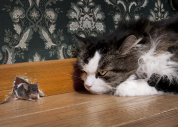


Hairballs are a constant problem in cats. A cats natural tendency to groom itself leads to hairball production. Causes are natural grooming and any other physical condition that causes hypergrooming such as flea bite dermatitis, eosinophilic complex or food allergies. Long haired cats have more severe hairball issues due to their thick long hair-coats.
During grooming, hair is swallowed and ends up in the stomach or the beginning segment of the small intestine. Hair is a natural irritant to the gastrointestinal tract. In some cases the hairball sits in the intestinal tract causing off and on vomiting. Many hairballs will pass through the cat and pass in the feces. Some may become lodged in the colon and become as hard as concrete. These are called trichobezoars and they can easily obstruct a cats lower colon.
The most commonly seen clinical sign in cats is off and on vomiting. Some days all will be right, another day the cat will vomit. The consistency of the vomit differs from that of a viral producing gastroenteritis. In hairballs, the food is vomited almost like it just was swallowed; totally undigested. It is very difficult to reproduce in writing, but the cat will make a characteristic sound during the whole exercise of vomiting the hairball. Cats will often sit for hours trying to get a hairball out of its system. Many of those than vomited will have a cigar like appearance. These are hairballs that were refluxed back into the stomach and than vomited to the outside.
Radiographs or a barium series may be performed if there is a suspect hairball obstruction. This will localize the location of the trichobezoar. Hairballs are one of the multitude of medical causes of vomiting in cats. Other tests such as a CBC and Chemistry profile may be done to rule out other issues.
Diagnosis of hairballs is by finding vomited hairballs or in many cases, hearing the characteristic heave ho sound made by a cat prior to vomiting the hairball. Radiographs are used to diagnose hairballs lodged in the lower gastrointestinal tract.
Treatment is by lubricating the gastrointestinal tract so that the hairball passes unobstructed into the feces. Many preparations are available. One of those is Laxatone®. Do not use petroleum jelly. Over time, it will bind fat soluble vitamins (D, E, K and A) causing a deficiency in them. The majority of hairball preparations available are fortified in these vitamins. The easiest way to get a cat to ingest the product is to put some on the paws of the front feet. Most cats hate anything stuck on them and will lick it off. Using a finger or tongue depressor, rub some on the roof of the cats mouth. The animal will act like it is gagging but the product will melt and the cat will swallow it. Hill’s® Science Diet Adult Mature Cat Hairball Control diet is also employed to slow down the development of hairballs.
Treatment of trichobezoars in the colon are sometimes relieved by enemas. If that fails, surgical intervention takes place and the obstruction is relieved.
Prevention also plays a big part in hairball control. All cats should be brushed and combed on a regular basis. If the cat will tolerate it, shampooing will also rid the cat of loose hairs and dander.
The prognosis for hairballs is excellent for most cats. Hairballs are just a fact of life for felines. Prevention by periodic hairball remedies and daily grooming will go a long way in keeping hairballs out of your hair!

