Common Diseases & Conditions in Pets B-C

Bladder stones are caused by the initial presence of struvite, oxalate or uric acid crystals in the urine. Over time they snow ball and become larger. They are ultra common in all Miniature Schnauzers, Bichon Frise amongst others. In cats, bladder stones form a component of Feline Urological Syndrome (FUS).
In dogs and cats, struvite crystals are the most common crystal found in the urine. Struvite crystals coalesce to form magnesium phosphate stones. Oxalate crystals form oxalate stones and uric acid crystals form urate stones. Urate stones are most common in Dalmatian dogs. Rather than secrete urea like all other mammals, Dalmatian dogs secrete uric acid (like chickens) and are at a high risk of urate stone production. In the presence of a urinary tract infection, organisms such as E. Coli split urea into various components causing the pH of the urine to rise. This alkaline environment favors the formation of urinary calculi (stones). Once formed, stones irritate the bladder lining causing blood in the urine. The bladder volume decreases making the animal want to urinate more frequently. Bladder stones hurt and cause the animal to strain to urinate. Male dogs and male cats have a narrow urethra. Small stones can block the outflow of urine and cause a urethral obstruction. Obstructions never happen in most females.
Sometimes the only clinical sign observed by owners is the periodic presence of blood in the urine. This is the most common sign presented to clinicians. The same thing happens with cats. Upon further questioning, many dogs are urinating more frequently and straining to do so. Sometimes the owner reports zero clinical signs. I once had to spay a dachshund and noticed a huge bump on the skin in the lower abdomen while it was on its back getting prepared for surgery. On palpation; I could feel a huge bladder stone.
A urinalysis is the order of the day here. It will not only give the doctor specifics regarding cells and urine pH but many times visible crystals will be seen that are easy to diagnose. Once the bladder calculi are removed, stone samples are sent off to the lab for definitive diagnosis. This facilitates which diet to put the animal on post op or after stone dissolution. In suspect cases, radiographs are taken (or ultrasound) which will demonstrate the presence of calculi in the urinary bladder.
Treatment of urinary calculi in the dog is usually done by surgical excision. This is called a cystotomy. The stones are removed and sent to the lab for definitive diagnosis. If there is only one or several stones, veterinarians may opt for dietary dissolution of the stones. The most common diet is Hill’s® Prescription canine s/d. Long term use of this diet is not recommended for over 6 months. This diet is also restricted in Magnesium; one of the components of struvite (Magnesium Phosphate) stones. Dogs will be put on antibiotics and a urinary culture may be performed. Once the stones are removed and or via special diet, dogs are put on a special urinary tract diet such as Hill’s® Prescription canine c/d for long term control. Periodic urinalyses are recommended to stay on top of the condition. Stones may recur though and many animals have to undergo several cystotomy procedures over their lifetimes.
In cats, surgical removal is rarely done unless the bladder is full of struvite grit. The big problem with male cats is that they may obstruct. This is a medical emergency and the animal is anesthetized and the obstruction is relieved by effective catheterization using a tom cat catheter. The grit is sent away for chemical analysis. Dissolution of feline struvites stones may also be dissolved via Hill’s® Prescription feline s/d. This diet should not be fed for longer than 6 months. After the stones have been dissolved, the cat has to be maintained forever on Hill’s® Prescription feline c/d or Royal Canin feline SO diet. Even if cats are off of this diet for just a few days, signs of cystitis can start up again. Veterinarians may also prescribe glucosamine/chondrointin which is often used to treat idiopathic cystitis in cats as well as arthritis.
The prognosis for urinary calculi in dogs and cats is excellent as long as medical care is sought in a timely manner and the owner continues to follow up with periodic urinalyses plus the appropriate urinary diet.


Blindness is defined as the inability or only partial ability to see from either one or both eyes. The causes of blindness are many. Congenital causes such as animals born with toxoplasmosis may demonstrate a retinopathy that inhibits vision. Older animals and diabetics develop cataracts that can lead to blindness. Glaucoma can lead to blindness. Eye trauma, such as ocular proptosis, can also lead to blindness. Proptosis is when the eyeball is forced out of its eye socket. Chronic corneal ulcers, chronic pannus in the German Shepherd and dry eye are leading causes of blindness.
Vision is a very complex wonder. The front of the eye and the iris physically see a flower. That flower image is transmitted through the lens to the retina in the left eye. From the retina it passes through an optic nerve. The same thing happens in the right eye. Those nerve impulses in the optic nerve cross at the optic nerve chasm behind the eyes. Those sensory nerves connect to the cortex and than via other motor neurons the impulses are than sent back to the eye where the image is than visualized. Any disease process that interferes with any of these steps may lead to blindness.
Regardless of the cause, vision loss is easily detected often by the actions of the dog or cat in the home environment. Both dogs and cats may bump into objects, become disoriented outside or inside, become startled when pet or touched, reluctant to jump on sofas and beds, unable to find the door to go out, inability to find its food and water bowls. Cats are very agile creatures. They may jump up on an object they previously did with ease but miss the mark and fall down. Owners often report that they see “something milky colored” in the eye; referring to cataracts.
Prognosis on blindness is proportionate to the rapidity at which the causing agent of blindness is diagnosed. Prevention is key. Medical care must be immediately sought after ANY eye injury. There is something positive though. Blindness does not inhibit a good quality of life in dogs or cats. Dogs can only visualize a few colors and greys plus are near sighted their entire lives. Both cats and dogs depend on their ability to smell. This is far superior to a human’s ability. This sensory ability allows, with a little bit of human help & TLC, to live a satisfying life.


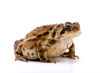
The Bufo toad is an animal seen throughout Florida and other subtropical areas. It is most commonly seen during the long summer rainy season; usually at night or early evening hours.
Bufo toads are active in the evenings and dogs see them jumping around and are fascinated by them. They chase after them and get the toad in their mouths and often eat the creature. The toad secretes a potent neurotoxin on its skin which causes seizures. The effects are even worse if the animal ingests the toad.
The most common clinical signs noticed are: drooling and salivating excessively, lethargy, staggering and seizure activity that will not resolve on its own. Smaller dogs are more prone to the toxic effects compared to a large breed of dog.
Bufo poisoning is a medical emergency. Veterinarians often do not get a history of bufo involvement unless the owner reports seeing the dog with the toad. Therefore a CBC and Chemistry profile are immediately drawn for study.
Diagnosis is readily made if the owner actually sees the toad in the dogs mouth. If the time of year is the hot, rainy season and a seizuring dog is in front of you Bufo poisoning is at the top of your medical rule out list. To get a doctor on the case STAT, they are called “frog dogs” in emergency clinics.
Owners at home are the first line of defense. They should carefully rinse out the mouth of the dog with fresh water yet prevent the animal from swallowing it. Bufo poisonings are treated like any other seizuring patient. They are hospitalized and giving convulsant therapy such as diazepam, pentobarbital or an inhalant anesthetic to bring the seizuring under control. Other supportive care measures such as an intravenous line with fluids and dextrose maintain body functions until seizure activity stops. Most dogs are discharged in a day or so with no further seizuring IF the seizuring was actually caused by the Bufo Toad.
The prognosis for Bufo Toad poisoning is excellent if it is recognized early and the animal receives appropriate treatment. Problem is, is that dogs are presented multiple times for said poisoning. Prevention is key. Keep the pet on a leash at night or indoors all the time; particularly during rainy, humid summer nights.


A bone fracture is a break in any bone in the body. Fractures can occur anywhere and are caused by many different means. The most common type of fractures seen are those produced by trauma. This includes car accidents and fights with other animals. Other fractures can be pathologic, that is caused by a disease process. Osteosarcoma is an example of this type of fracture. Fractures can most easily occur in tiny breeds such as the Maltese. Just dropping them from your arms on to a tile floor can cause fractures. Cats suffer from “high rise” syndrome. They can survive relatively falls from medium height by twisting and turning but often land wrong causing head and pelvic fractures. Fractures are classified as open or closed. Open fractures are those that penetrate through the skin and can be seen. Closed fractures are those that do not penetrate the skin surface.
As an animal matures, bones become calcified and are more prone to fractures than young animals. Young animal bones are more flexible and often do not cause a break compared to a mature dog going through the same trauma experience. These are called greenstick fractures. Depending on the amount and direction of the force, fractures can occur in many different forms. Some are sheared off perpendicular (transverse), others form a spiral pattern yet many are obliterated and have no remaining blood supply to the bone.
Bone Fractures are characterized by pain, swelling and lameness on the effected limb. Fractures can mimic other medical conditions such as a bite wounds, muscle tears or soreness and hip dislocations. Additional signs are seen depending on the fracture location. Fractures of the lumbar vertebrae may cause paralysis of the hindquarters and inability to hold urine.
The most important lab tool is to radiograph the suspected fracture site. This is crucial. Many fractures are trauma induced so veterinarians will do a CBC and Chemistry profile to rule out other issues. Since all fractures are repaired under a general anesthetic, having blood work available prior to surgery insures it is safe to anesthesize the animal.
Diagnosis of a fracture is made from a good history, physical exam and radiographs.
The initial treatment of a suspected fracture should start at home. It is ultra important to be careful how the animal is handled. It is wise to put a cloth muzzle on the animal. It may bite otherwise. This is not out of anger but is born out of acute pain! Gently attach a newspaper or magazine around the effected area and gently tape it. It is much easier at this point to pick the dog or cat up so that the fractured area is not touched while moving the pet. In the car, try to initially position the animal by placing it on the OPPOSITE side of the fracture site. Once at the hospital, veterinarians will diagnose than treat the condition. If the animal was hit by a car, that trauma is taken care of first so that the animal is stable than the fracture repair can be addressed once the pet is a good anesthetic risk. Fractures can be repaired by: direct reduction, bone pinnings, plus metal plates and screws. External fixation devices such as casts and splints are commonly employed. Cats are superb healers. Many suffer from severe pelvic fractures. In 8 weeks, without doing anything, they heal like nothing has happened to them! However, the female must be spayed to prevent obstetrical complications due to a small birth canal caused by the pelvic fracture. Post reduction, animals are put on and sent home on painkillers, anti-inflammatory drugs and antibiotics. Other medications depend upon the actual cause of the bone fracture.
The prognosis for fractures depends upon the type of fracture and how long it took for the animal to receive medical attention. Fractures in young, healthy dogs have a very good prognosis; normally healing in about 8 weeks. Fractures that have a poor bone supply may not heal at all. These are called non union fractures. Fractures caused by cancers such as osteosarcomas have a poor prognosis. Prognosis is variable in all cases.


The prostate gland is an accessory sex organ in the male animal whose main function is to contribute fluids to semen. It is located behind the bladder close to the neck of that organ. The outflow tract of urine from the bladder goes through the middle of the prostate as it courses to the outside as the animal urinates. BPH is an enlargement of the prostate seen in UN-NEUTERED male dogs. Enlargement is under hormonal control (testosterone). Most non neutered male dogs over the age of 5 have an enlarged prostate gland. Prostate bacterial infections, adenomas, cysts and hyperplasia are possible but the most common is BPH due to testosterone.
When the prostate gland enlarges certain things happen. The gland grows upward putting pressure on the rectum making it harder for the animal to defecate. This is known as tenesmus. As the gland enlarges it puts pressure on the urethra compressing the diameter of it making it harder for the dog to urinate. In some cases this narrowing of the urethral lumen (opening) can cause a complete urethral obstruction. This is a medical emergency.
The most common clinical signs noticed are un neutered dogs that are straining to urinate or defecate. Some dogs will have a blood tinged urine (hematuria) or speckles of bright red blood in the stool (melena).
Radiographs or other imaging devices may be employed to visualize the prostate gland. To rule out bacterial prostate issues amongst others, a CBC and Chemistry profile are routinely performed. A urinalysis is always done.
Diagnosis is made by the history of an un neutered middle aged male dog (and up) plus clinical signs. Physical exam confirms a diagnosis by rectal palpation of the prostate. In most dogs with prostate enlargement, the prostate can be palpated yet difficult to touch as the gland grows and hangs over the cranial pelvic rim. Lab Work also aids in the diagnosis.
Treatment of prostate enlargement depends upon the cause of the enlargement. Infections require long periods of antibiotics as it is very difficult for antibacterials to reach the prostate gland itself. In hormonal enlargement, most dogs are neutered and the prostate gland usually shrinks back to normal in 2-4 weeks.
Prognosis for most prostatic enlargements is fairly good once the animal has been neutered or on long term doses of antibiotics for prostatitis. Prostate cancers, at the moment, carry a poor prognosis. I have only diagnosed one in my career. Prevention is key here. To avoid prostate enlargements, all animals should be neutered at 6 months of age. If used for breeding, neutering should be performed after stud services are complete.


Bracheocephalism is a condition, not a disease. It is a type of skull structure seen in Boston Terriers, English Bulldogs, Pugs, Persian cats and others. The face has a sort of smushed up appearance and this causes the sinus passages to run tortuous throughout the head. Eyes may appear to extend out more from their sockets (exopthalmic). The heads tend to be rounder than other breeds. The cause is genetic. To maintain a certain breed requires a set of genes that are expressed as the phenotype (appearance) of a particular breed.
Because of the skull structure, many of these breeds have many medical problems. Many are intolerant of heat and have difficulty breathing in those conditions. Because everything is so compact in the back of the throat (pharynx) excessive heat can cause the trachea (windpipe) to close. This is made worse as many of these breeds have an elongated soft palate. This condition is exacerbated if the animal is obese. Eyes tend to be overly exposed making them prone to dry eye and corneal ulcers. The nasal openings (nares) are narrower than normal making it harder to get oxygen into the lungs.
Most bracheocepalic breeds can live normal lives but their anatomical quirks can get them into trouble. Most problems deal with their respiratory tract. Dogs with a longer skull type may breeze through a simple respiratory problem. Not so with these dogs. They will snort, have tons of mucous secretions produced and have general difficulties getting air in and out of the lungs. They always are at a high risk of heat stroke. Their eyes may easily develop conjunctivitis and corneal ulcers that are characterized by conjunctival inflammation and scratches on the front of the eye.
Since most problems revolve around the respiratory system, most of these animals should be radiographed plus a CBC and a Chemistry profile performed. Cultures may be obtained to accurately diagnose the cause of infection.
Diagnosis is made by recognizing all the bracheocephalic breeds and medical problems associated with them. A medical history and exam plus lab data will point the veterinarian in the right direction. The goal is to find an appropriate treatment.
Accurately diagnosing a bracheocephalic dog while it is wide awake is extremely difficult. Not only is the oral cavity small but the tongue is short and thick. Many times the animal has to be sedated to find the exact cause so as to decide on a treatment. This is especially so with elongated palates. Bracheocephalic breeds that have off and on respiratory problems during excitation, excess heat or other causes are prime candidates for soft palate resection. This is usually done via laser surgery. Surgical opening of the nares is another procedure. I have done this on Persian cats. It definitely makes breathing easier. If the animal is obese, it has to lose weight, no ifs or buts. Excess weight makes clinical signs worse.
The outcome or prognosis for most bracheocephalic dogs that undergo soft palate resections, nares enlargement and weight control (if obese) is good. Those dogs that have chronic ailments such as pulmonary or laryngeal disease are at a disadvantage and hence have a much more guarded prognosis.


Female dogs and cats have five pairs of breast. Mammary glands are needed to produce milk to feed nursing newborns. The cause of the majority of breast cancers is estrogen excess. Dogs and cats that develop false pregnancies or irregular estrous cycles are also at a higher risk of developing breast tumors. Small breeds such as: Maltese, Yorkshire Terriers, Toy Poodles and Chihuahuas are at a higher risk of developing breast tumors.
Female dogs and cats that are spayed before their first heat cycle have an almost zero percentage chance of developing breast cancer. Each heat cycle that a female animal goes thorugh increases the chance of developing breast cancer by about 8%. The majority of breast tumors in cats are malignant. Those in the dog are mixed; meaning about half of them are benign adenomas while the others are malignant and can metastasize to the lungs, lymph nodes and liver.
Clinical signs are associated with firm breast masses that occur from the first pair of breasts down to the fifth pair. Tumors that can easily be moved around are usually benign while those that are fixed to the subcutaneous tissues below it are often malignant. Mammary tumors in dogs and cats are most commonly seen in the fourth and fifth pair of glands in the groin or inguinal area. These glands produce much more milk and are much more active than the first three pair.
Chest films are always taken to see if there is any metastasis of the mammary tumor to the lungs. CBC and Chemistry profiles are also run to detect mammary activity elsewhere.
Diagnosis is made by the presence of an intact middle aged female dog that has one or several firm masses over the mammary gland area. Some animals were spayed at an earlier age but after 6 months of age. The actual pathological diagnosis is made by submitted excised mammary gland tissue to determine the exact cellular diagnosis of the mammary tumor.
If the animal may be safely anesthetized, the best treatment is the surgical removal of the mammary gland and mass with or without the removal of associated lymph nodes. I was taught to always spay the dog at the same time to prevent further mammary tumors from developing. Now a days this is controversial. This still makes sense though, since spaying the animal at that time will prevent pyometra, hydrometra and other gynecological diseases from happening. While in the abdomen, it is a good idea to look at the liver lobes and any other organs for any metastatic activity. Some animals, at specialty practices, may receive radiation and other care. If infections are taken care of and seromas are prevented, most dogs and cats do fine post surgery.
The prognosis for breast tumors depends upon the histological diagnosis of the tumors. Complete excision of a benign tumor receives a favorable prognosis while the excision of a malignant tumor, with or without metastasis, receives a much guarded prognosis. The best course is prevention. All female dogs and cats should be spayed prior to their first estrous cycle. Those that are used for breeding should be spayed at about 3-4 years of age. Those individuals that find or become owners of an intact adult female should immediately have her spayed.


The bronchi are the tubes that carry oxygen to the alveolus; the end of the bronchioles where oxygen is exchanged for carbon dioxide. They bifurcate into left and right bronchi. Off of each of these are smaller air tubes. Bronchitis is the inflammation of the tubes; the bronchi. Infections can be acute or more commonly chronic. Causative agents are: bacterial, virus, inhalant allergies, presence of smoke or any other irritant. Any dog or cat can get bronchitis but those that suffer the most are those bracheocephalic dogs (see preceding disease condition) such as the Pug, Persian cat or the English Bulldog.
The hallmark of bronchial disease is inflammation. Inflammation causes excessive production of mucoid secretions. These secretions can occur up and down the bronchial tubes. They clog the airways making it more difficult to breathe. In chronic cases of bronchitis, the lumen (diameter of any hollow organ) of the bronchi decreases. This becomes a vicious circle making it harder and harder for the animal to get oxygen where it is needed. This mechanism is what explains the majority of clinical signs.
The most commonly seen sign associated with acute and chronic bronchitis is a dry, raspy cough that is exacerbated by any type of exercise or excitement. The cough may be productive (coughing up phlegm) or non-productive. Depending on the cause, some dogs will run a low to moderate fever. Excessive exercise makes it difficult to breathe so the respiratory rate will increase and the animal finds itself straining to catch its breath (dyspnea). In severe cases, the dog will have excessive abdominal respiration; meaning very little inspiratory ability of the lung tissue. Animals are also anorexic and are off their food due to discomfort or just being too tired to get to a food bowl.
Lab work will help to identify the cause of the bronchitis. A CBC and Chemistry profile are performed and 2 chest films are taken via standard radiography or other imaging techniques. Radiographs will show excessive bronchiolar infiltration. Cultures of the phlegm or a tracheal wash may be done to identify any infectious agent.
Diagnosis is made by obtaining a solid history and a complete physical exam. Auscultation of the chest will produce sounds of wheezing and moist or dry rales over the chest. Palpating the trachea via the skin will usually elicit a dry hacky cough. The cough heard usually sounds like a goose! Because of the difficulty breathing, the animal abducts (extends its forelimbs) to make it easier for inspiration. Combining the exam and history with lab work makes it pretty straight forward to figure out.
Treatment is based on treating the cause of bronchitis and managing the clinical signs of the disease. Most cases will require antibiotics to treat infectious bronchitis or prevent a secondary bacterial infection in other cases. Allergic cases are treated with corticosteroids or inhalers. Nebulization is one of the best treatments for these animals that make both dogs and cats feel and breathe so much better. This should be done at least 4-5 times per day initially. The nebulization therapy involves inhaling: Mucomyst®, aminophylline, gentomycin and dexamethasone. If the cough is non-productive antitussives such as butorphanol are used. This has to be given often but it also takes away the pain and encourages the animal to rest. Bronchodilators such as theophylline are used to open up the airways to allow as much air in as possible. Pets are sent home on a combination of these drugs. Chronic cases can be difficult to treat and involve cough suppresants such as Hycodan® plus non-systemic corticosteroids such as budesonide. Home supportive care is crucial. Animals should remain in an air conditioned environment and air handler filters changed more frequently. Short walks are okay but use a harnass rather than a leash and collar. This will decrease inflammation and hence the cough reflex.
The prognosis for acute bronchitis is usually good, as long as the owner seeks immediate veterinary care and appropriate therapy is started. The prognosis for chronic bronchitis is guarded in that many of these animals enter a downward spiral where medical care does very little for the animal; even breathing pure oxygen. On the other hand, prior to this, many of these animals can be maintained successfully on medical therapy and corticosteroid inhalers to enjoy a relative good quality of life.


Burn wounds are extremely painful, not only for humans, but for our companion animals. The cause of burns are usually broken down into three groups: heat (themal), chemical and radiation. Thermal causes are the most common and occur in the home the majority of the time. Cats jumping up onto a turned on electric stove or a woodburner in full blaze will suffer burn wounds to the pads. Hot liquids, lamps and excessive use of heating pads can cause burns. Burns are also produced by caustic chemicals such as products containing acids. Radiation causes include those dogs undergoing radiation therapy. In my Ohio practice I had to treat a cat that was put in a microwave oven by a little boy for about half a minute. The cat did survive. Dogs also get into trouble (particularly puppies during teething) by chewing on electrical cords. These produce burns on the mouth or gum tissue.
Regardless of the cause of the burn, cellular damage occurs. Tissues are composed of individual cells and these cells are literally cooked and undergo lysis and when grouped together, tissue damage. This is the most important consideration of burn pathophysiology in a nutshell.
The severity of burns is grouped into three types. First degree burns are those that effect the epidermis or top skin layer. These occur during mild sunburn. Dogs and cats without hair such as the Chinese Crested dog and Sphinx cat are susceptible. Secondary degree burns are those that cause damage down to the dermis or deeper layer. These burns are associated with blistering and require medical care. Third degree burns destroy and char the entire skin surface. The tissue is dead, circulation damaged and the body is prone to bacterial infections. These are dangerous burns.
All burn patients should have a CBC and Chemistry profile done numerous times while being treated for burn wounds; particularly those animals being treated for third degree wounds.
Diagnosis of a burn wound is pretty easy if the burn is noticed by the owner. Some burns may show themselves as time goes on and or be hidden by the animal’s hair-coat.
Initial first aid may begin at home. Cold compresses can be applied to thermal wounds such as sunburn. Chemical burns should be flushed with running water to drain off as much contaminant as possible. All burns patients should than receive medical care. The treatment is based on which degree of burn is present, the location of the burn and what percentage of the body is being effected. Blood is drawn to check organ function and an intravenous line is installed with fluid therapy. Burn patients suffer from dehydration. All burn patients (particularly third degree) suffer from intense bacterial infections caused by Pseuodomonas aeringinosa. Intravenous and oral antibiotics are ordered. Surgery is required in third degree burns. The goal is to debribe as much dead tissue away as possible and attempt wound closure. Many animals require grafts but if this is not possible, burn wounds will heal and fill in with scar tissue. Scar tissue does not have hair follicles so the area will be devoid of a hair-coat.
The prognosis for burn patients depends upon the degree of burn and the percentage of the skin surface that has been burned. Primary burns have an excellent prognosis while severe burns that cover 30%-50% of the body surface have a poor prognosis. Antibiotic and fluid therapy are required for a long period of time. Wound healing takes a long time. Best bet is prevention of burns by keeping caustic chemicals away from pets, minimize exposure to the suns ultraviolet rays and do your best to isolate dogs from electrical cord chewing. Trying to keep cats from jumping is futile so if you own a cat always stand by the stove when cooking.


Brucellosis is caused by the bacteria, Brucella canis. This organism is a Gram positive rod that causes reproductive issues and fertility problems in male and female dogs.
Brucella canis is difficult to deal with. Transmission of the disease is during sexual contact during mating season. Since a lot of these dogs have abortions or stillborns, dead puppies are ingested by the mother. This is normal behavior for the bitch but can also transmit the disease. This organism hides out in certain cells in the blood making it difficult to treat. Brucellosis is a zoonotic disease, meaning it can be transmitted to humans. Care must be taken to prevent human exposure.
The classical clinical signs associated with Canine brucellosis are abortions in the third trimester (last 20 days of pregnancy) and future infertility in the male or female animal. Stillborns are also very common. In the male, the penis may be infected as well as the prostate gland. Some animals are also asymptomatic.
Both male and female dog used for breeding should undergo a Brucellosis test. This is extremely important in preventing transmission back and forth.
Diagnosing brucellosis is performed by sending a blood sample to a lab that performs an agglutination test. If it is negative your dog does not have brucellosis. However, there may be false positives so a positive test result should prompt the veterinarian to confirm the diagnosis by other means such as culture of stillborns. As with Babesiosis, a PCR test can be used to diagnose the disease.
There is no effective, complete treatment of the disease. Antibiotics such as doxycycline, over a long period (and others), have been used. This is ineffective in the long run as animals will still have Brucella organisms hiding from the immune system and antibiotics while hiding in certain cells like macrophages. There is little else to be done. Spaying or neutering the animal prevents future sexual transmission but does nothing for the organisms already present. Often euthanasia is performed in these cases.
The prognosis for Brucellosis is poor in most animals. All treatments are ineffective at eliminating the bacterial infection. Care must be taken to minimize human exposure to secretions and body parts associated with Brucellosis. This means gloves plus eye and nose protection.


Calicivirus is one of the most common causes of respiratory disease in cats; particularly in young kittens. The virus itself, has numerous serological types and these differ in the severity of disease presented in the cat.
Calicivirus is extremely infectious to other cats. It is most common where cats are found the most in close quarters. This includes boarding facilities and animal shelters. The virus incubates in the cat for about 3 or so days. The infection lasts for about 3 weeks but during that entire time, the cat can transmit the disease to other cats. Other cats that cause problems are carriers. These are clinically normal cats that carry the virus and can easily infect other susceptible cats. Pregnant queens can transmit the disease in utero to her unborn kittens. This is a nasty feline virus and easily prevented. It is a member of the core feline vaccination program.
Clinical signs of calicivirus vary depending upon the serological strain that the animal is infected with. Most clinical signs are respiratory. A bilateral muco-purulent nasal discharge is present forcing the cat to sneeze a lot. A hallmark of calicivirus are the oral ulcers that are found throughout the oral cavity. Many cats also will have bilateral conjunctivitis which is also painful. Cats are also lethargic and have very little appetite. Cats have to smell their food prior to eating it and the olfactory device ie: the nose can not perform that job well when sick.
All viruses cause a drop in white cells so a CBC is performed to evaluate the cellular immune response ie: by looking at the neutrophil and lymhocyte counts. In certain cases veterinarians can have the virus diagnosed via a PCR lab test.
Diagnosis is usually made by the oral ulcers present in the mouth cavity plus the majority of patients are usually 6 months of age or younger. It can also be diagnosed by a PCR lab test.
There are no antibiotics that effectively treat any virus. Viruses lower the immune response making the animal susceptible to a secondary bacterial infection. This is why most veterinarians will put the animal on an antibiotic such as Clavamox® drops for 10 days. An opthalmic ointment is also dispensed to take care of cats that have conjunctivitis. Supportive care such as nebulization/steam vapor will make the cat feel more comfortable. It is important that animals start to eat as soon as possible and I highly recommend Hills® Prescription a/d for this purpose.
With appropriate medical care the prognosis for calicivirus patients is excellent. The only problem is that they are highly contagious for 3 weeks to other susceptible animals. The best bet is prevention. This means starting kitten vaccination at 6-8 weeks, followed by a booster in 4-5 weeks than once annually.


The cause of canine hip dysplasia is a faulty development (in congenital cases) of the hip joint and in middle aged to older dogs, a breakdown in joint cartilage leading bone to grind against bone causing intense hip pain and arthritis. It is a genetic disease in German Shepherd dogs but is most commonly seen in large breeds of dogs such as: Rottweiller, Great Dane, Golden Retrievers and Labrador Retrievers.
In congenital Canine Hip Dysplasia the angles of the femur and pelvis are not normal and this causes abnormal wear and tear on the hip joint ie: the femoral head and acetabulum. This leads to clinical signs. In middle aged dogs and up, bone cartilage degrades allowing bone to grind against bone causing inflammation and the resultant lameness and discomfort.
The most common clinical signs are limping on the affected limb, reluctant to exercise and a reluctance to go upstairs. Animals may cry out when touched or pet on the hind quarters. This is known as referred pain. If left untreated, dogs are immobilized and are unable to get up to urinate or defecate or even get to the food bowl. These are called downer dogs and are usually seen in advanced cases.
The most effective lab tool is radiographing the hips. This entails two different views at perpendicular angles. The degree and or severity of the dysplasia is easily figured out. If an animal is older, a CBC and Chemistry profile are in order. Clinical signs of hip dysplasia can mimic other disease processes such as fractures, osteosarcomas and muscular dysplasia in the quadriceps. For breeding purpose the Orthopedic Foundation for Animals (OFA) offers a diagnostic test to ascertain if animals should be bred. Dogs that test positive should not be bred as the gene will be passed down to its progeny.
Diagnosis in lame animals is made via a good history and physical exam. Radiographs will confirm a diagnosis. In healthy animals, radiographs interpreted by the OFA will diagnose the condition. The dogs need to be at least two years of age for OFA certification.
Treatment of Canine Hip Dysplasia is usually directed to correcting as many of the arthritis problems present as well as controlling pain. In advanced cases, animals may undergo a complete hip replacement at specialty practices. The majority are treated medically. Joint inflammation is taken care of by special anti-inflammatories such as Rimadyl®. Maintenance on this drug requires periodic liver tests. Some animals are so severe that they require corticosteroids. Pain can indirectly be managed by Rimadyl® or by a pain killer such as Tramadol®. The joint cartilage can be rebuilt in many cases, if started early enough, by taking glucosamine/chondroitin everyday. These dietary supplements take about 3 weeks to take effect but the results in many cases can be dramatic. The skeletal system alone does not just support the animal. Muscles also contribute so it is important, if possible, that the animal is gently exercised to maintain muscle tone. Many animals suffering from hip dysplasia are overweight. This puts too much pressure on effected joints. A weight loss program is essential. Other animals are obese due to hypothyroidism. Several years ago, I had such a case in a Golden Retriever. All older dogs should have thyroid tests performed (T4, FT4). Supportive care at home is important. Provide thick, comfortable bedding for the dog. Care must be taken so the animal does not injury itself on slippery tile or wood floors. Massaging the muscles in the effected leg may help. Having practiced up north for many years, I know for a fact that these dogs suffer more in cold, damp weather. That is a good enough excuse to move to the sun belt!
The prognosis for dogs depends upon the severity of the dysplasia and how long the dysplasia has been present. Dogs diagnosed early and treated right after, have a favorable prognosis even though the condition can not be cured. Dogs that are unable to stand up due to chronic degenerative dysplasia have a poor prognosis.


Canine Influenza virus (Canine Flu) has been infecting many dogs over the last 4-5 years. Canine Influenza is caused by the type A virus and exists in two different forms. All flu viruses are caused by myxoviruses; a RNA type virus. The canine virus originated initially from horses.
There is no known transmission of canine influenza to humans but officials have to be alert. The fact is, is that it mutated from horses to dogs. Myxoviruses (Canine Influenza) are chameleons when it comes to their antigenic traits. They are changing all the time. This is caused by antigenic shift and drift. This is why vaccines often do not work year to year as the surface antigens change all the time. Because of this mutation, exposure to humans could eventually be possible; just not yet at this time. Canine Influenza produces signs of fever and respiratory illness in some yet some animals do not show any signs but can be carriers of the disease. Some animals develop severe disease which is usually associated with pneumonia. As in humans, canine influenza has a high morbidity rate yet low mortality rate.
The most common signs associated with canine influenza are: fever, runny nose and cough plus general malaise. Lethargy and anorexia are common. Some animals do not show any clinical signs yet others can become severely ill with pneumonia.
There is a test to diagnose canine influenza that was recently put out by Idexx Laboratories. Animals that are presented with influenza require a CBC and Chemistry Profile. Radiographs need to be performed to check pulmonary function and rule out pneumonia.
A Diagnosis is made by clinical signs and history. The latter is extremely important. Dogs that are housed in close quarters, such as at groomers, boarding kennels and animal shelters are at a much higher risk of getting canine influenza compared to a single pet household. Diagnositic radiographs and blood work help guide the doctor to a diagnosis.
There is no direct treatment for canine influenza. All medical care is supportive. This insures maintaining fluid needs, caloric needs plus relieving respiratory discomfort with steam vapor or nebulization therapy. Most animals will also be placed on an appropriate broad spectrum antibiotic to prevent secondary bacterial infections. Animals that suffer from pneumonia are hospitalized, treated and radiographed periodically to hopefully see a clearing of the lung fields. The ability to clear the influenza virus depends upon the strength of the dogs individual cellular immunity plus humoral immunity that leads to active antibody production to the virus. Animals that are immunosuppressed (cancers, autoimmune diseases, Cushing’s Syndrome, Diabetes mellitus) may have difficulties mounting a successful immune response.
The prognosis for most cases of Canine Influenza is favorable. Although there is a high morbidity (meaning many animals are susceptible) there is overall a low mortality rate plus some dogs do not show any clinical signs at all. Prevention is becoming part of the game. There is a Canine Influenza vaccine that protects against one serotype of the virus (H3N8). Whether it neutralizes the other serotype (H3N2) is unknown. Animals that should be vaccinated are those that are around other dogs frequently. This includes dogs that are boarded or groomed often. A good rule of thumb is that if you currently have your pet vaccinated against kennel cough (Bordetella bronchiseptica), it is a good idea to have the pet vaccinated for Canine Influenza.


The actual cause of cardiomyopathy is unknown. It is most commonly seen in large breeds of dogs with the Doberman Pinscher being at the top of the list. Great Pyrenees, Golden Retrievers, Irish Wolfhounds are just some of the breeds afflicted with this. Any injury to the heart muscle could lead to cardiomyopathy. Ischemia (decreased blood flow), toxins, hyperkalemia (excessive potassium) and other agents could lead to heart muscle damage. In cats the majority of animals develop the condition from a lack of taurine or excess thyroid hormone production. Taurine is an amino acid that cats can not manufacture on their own and their diets must be supplemented with it. Excessive thyroid production (hyperthyroidism) is common in cats 10 years of age and up.
Cardiomyopathy can occur in two forms. Hypertrophic cardiomyopathy is a condition when the muscle of the left side of the heart thickens. This is very common in cats but not so in dogs. In dogs, the dilated version is more common which entails an enlargement and thinning of the left, right or both sides of the heart. This eventually leads to heart failure. The heart is unable to pump adequate amounts of blood. In left sided failure, fluid builds up in the lung and is known as pulmonary edema. In right sided heart failure the lungs do not get enough blood to oxygenate the red cells. Fluid can leach out, back up and cause edema in the neck and limbs. As the heart enlarges, the entire heart fails to work well. The enlargement puts pressure on the trachea causing a “heart cough”. If left untreated, there is complete oxygen depletion than can not support life.
Clinical signs associated with cardiomyopathy in dogs and cats are brought about by a failing heart. This is known as congestive heart failure. Animals may become tired and lose weight initially. Some actually have pathological arrythmias before actual signs develop. Shortness of breath and coughing develop. The animal can not get comfortable and will not exercise as it did once before. Even going up stairs can cause an animal to get winded or or turn cyanotic. In right sided failure fluid may accumulate in the abdomen (ascites) giving the pet a distended, pendulous abdomen. Cats are unable to jump up on objects as they did in the past. Sadly, in some cats there are no clinical signs but are found deceased by their owners often appearing like they are sleeping. Cats may also lose their vision since excessive blood pressure damages the retina.
A CBC and Chemistry Profile are extremely important. They will show any electrolyte abnormalities as well as liver or renal disease. Chest films and ultrasound are used to visualize the heart and surrounding lung tissue. Electrocardiograms are great to pick up any abnormal electrical activity such as arrythmias. One of the best diagnostic screening tests is the IDEXX Pro BNP test. This gives a quantitative interpretation of a dog or cats heart function. Values above a certain level are indicative of heart disease. It is an excellent tool to differentiate cardiac from pulmonary disease. In cats measuring taurine levels are useful.
Diagnosis is made by compiling a complete history and physical exam. Understanding the predilection of the disease in large breeds of dogs is helpful. Interpreting lab work (that was discussed right above the diagnosis) will lead the doctor to an appropriate diagnosis of cardiomyopathy.
The goal of treatment in dogs is to provide pharmaceutical support in order to strengthen the remaining heart muscle and to alleviate the clinical signs associated with the disease. Inotropic agents (drugs that strengthen the tone of heart muscle) like digitalis have been used but many veterinarians are using Vetmedin® (pimobendan) for that same purpose in dogs. It is not approved for felines. Diuretics such as furosemide and spironolactone are used to clear pulmonary edema. Ace (Angiotensin Converting Enzyme) inhibitors (benazepril) and beta blockers are often used to decrease return blood flow pressure to the heart.
In cats, the goal is to treat underlying thyroid and taurine disease. Clam juice is an excellent source of taurine. All cats above 10 years of age should be screened for hyperthyroidism. It will prevent a lot of cats coming down with cardiomyopathy in the first place as well as screening them with the Idexx® Pro BNP test. Once the underlying disease is being treated, alleviating the clinical signs of heart disease are important. The use of Calcium channel blockers, ace inhibitors or beta blockers plus furosemide are routinely used to treat heart failure in cats.
Dogs and cats should both be placed on a low sodium diet. Hill’s® Prescription Canine h/d is appropriate for dogs while this list provides adequate dietary control for cats.
The short term prognosis for dogs and cats treated for cardiomyopathy is pretty fair as long as the condition is diagnosed early with appropriate medical care and follow up visits. Regardless of the care, most animals eventually succumb to heart failure and are often euthanized when breathing becomes excruciatingly difficult due to excessive inflammation and untreatable end stage pulmonary edema.


Cat Scratch Fever is a condition seen in people and transmitted to the person by cats. The causative agent is Bartonella henselae.
Cats get infected from the bacteria by getting involved in cat fights and also from flea bites and flea dirt (feces). Bacteria is often found in the nail bed of the cat and is transmitted to other cats and people by means of scratching or biting the person.
Many cats are asymptomatic and act normal. Bite wounds may be simply treated medically without knowledge of a possible Cat Scratch Infection. In people, a bite or scratch wound becomes infected and peripheral lymph nodes closest to the wound become enlarged.
Most veterinarians perform a CBC and Chemistry Profile when diagnosing feline bites. It may point the veterinarian to consider other causes such as Cat Scratch Fever. Problem is, most cats are asymptomatic.
Diagnosis of bite wounds in cats is easy but figuring out if the cat is carrying Cat Scratch Fever is another thing. Most cases go undetected. For diagnosis in humans, consult your physician.
Treatment of bite wounds with Clavamox® and Baytril® may actually help clear Cat Scratch Fever in the cat. Most cats remain carriers so trying to treat something that is not diagnosed is difficult.
The prognosis for cat scratch fever in cats and humans is excellent. The best treatment is prevention. Young cats are the most active when it comes to scratching. They are using their claws to play and to learn how to hunt. People need to avoid playing with them during this stage. Cats should be put on topical flea control to kill fleas plus keeping cats inside with the goal of preventing transmission from fighting with other cats.

